**Experiencing persistent knee pain, clicking, or a feeling of your knee giving way can be alarming. These symptoms often point towards a potential injury to the meniscus, a crucial piece of cartilage in your knee joint. Understanding the nature of your knee pain is the first step towards recovery, and this often begins with a series of specialized physical examinations known as meniscus tests.** These diagnostic tools are vital for healthcare professionals to accurately assess the integrity of your menisci and guide you towards the most appropriate treatment. Every year, over 1 million Americans tear their meniscus, highlighting the prevalence of this injury. While an MRI is often used to confirm a diagnosis, clinical tests play a critical role in the initial evaluation, helping doctors narrow down the possibilities and understand the specific nature of your knee issue. This comprehensive guide will delve into the most common and effective meniscus tests, explaining how they work, what they look for, and why they are so important in the journey to knee health.
Understanding the Meniscus: The Knee's Vital Cushion
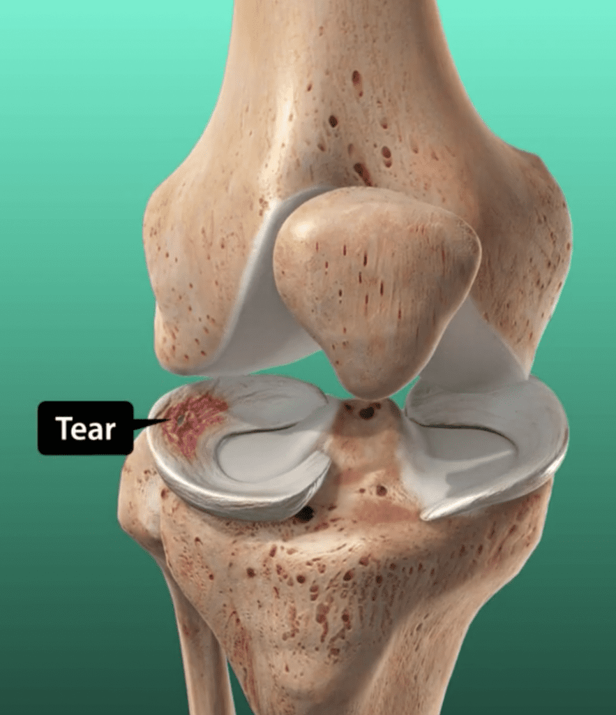
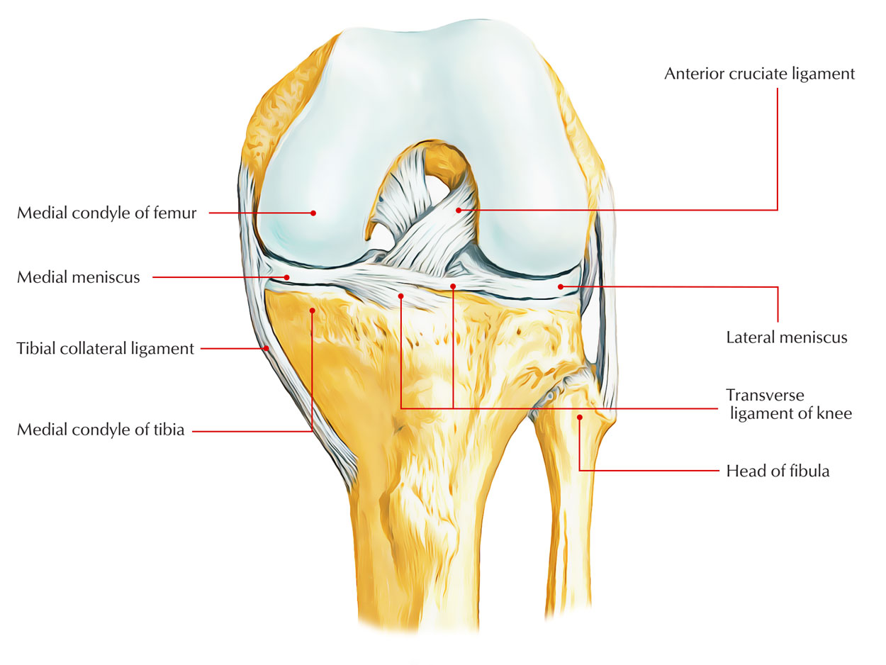
Before diving into the specifics of meniscus tests, it's essential to understand what the meniscus is and why it's so important. Your knee joint, one of the largest and most complex joints in your body, relies on two C-shaped pieces of cartilage called menisci. These are located between your thigh bone (femur) and your shin bone (tibia). You have two menisci in each knee: the medial meniscus (on the inside of your knee) and the lateral meniscus (on the outside). The medial and lateral menisci play an incredibly important role in absorbing force and assisting in the role of nourishing the knee joint. Think of them as shock absorbers, distributing the weight and stress across the knee joint during movement. They also contribute to knee stability, help lubricate the joint, and facilitate smooth movement. When these vital structures become injured, often due to twisting motions, direct impact, or even age-related degeneration, it can cause altered biomechanics of the knee joint and significant pain. Meniscal tears are the most common lesions followed by the meniscal cyst, indicating how frequently these injuries occur and the impact they can have on daily life.
The Importance of Meniscus Testing: Why Diagnosis Matters
Accurate diagnosis of a meniscal tear is paramount for effective treatment and long-term knee health. Without a proper diagnosis, a seemingly minor knee issue could escalate, leading to chronic pain, instability, and even further damage to the joint over time. This is where the various meniscus tests come into play. These physical examination techniques are designed to put specific stress on the menisci, provoking symptoms that indicate a tear. The goal of a meniscus test is to reproduce the patient's pain or symptoms, such as clicking, locking, or a sensation of the knee giving way. While these tests are not 100% conclusive on their own, they provide invaluable information to a healthcare provider, guiding them toward a presumptive diagnosis and informing the decision for further diagnostic imaging like an MRI. Early and accurate diagnosis means a quicker path to appropriate treatment, whether it's nonoperative methods like physical therapy or operative methods like surgical repair.
McMurray's Test: The Gold Standard for Meniscal Tears
The McMurray test, also known as the McMurray circumduction test, is arguably the most common orthopedic special test used in the physical diagnosis of meniscus lesions. It is a cornerstone of knee injury assessment, widely employed by doctors to evaluate individuals for tears in the meniscus of the knee. This test is designed to assess the integrity of both the medial and lateral meniscus by attempting to trap a torn piece of cartilage between the femur and tibia.
McMurray's Test: How It Works
Performing the McMurray test involves a series of knee and leg movements that can help diagnose a torn meniscus. The patient lies on their back while the examiner flexes the patient's knee fully. To test the medial meniscus, the examiner rotates the tibia laterally (outwards) and applies a valgus stress (force applied to the outside of the knee, pushing inwards) while slowly extending the knee. For the lateral meniscus, the examiner rotates the tibia medially (inwards) and applies a varus stress (force applied to the inside of the knee, pushing outwards) while extending the knee. The test involves extending and rotating the knee with varus or valgus stress and looking for pain, snapping, clicking or locking symptoms. The test involves applying varus or valgus force and tibial rotation while extending the knee. Depending on the meniscus we want to investigate, the patient’s feet are turned outwards (medial meniscus) or inwards (lateral meniscus). The anterior half of the meniscus is not as easily tested with the McMurray test, because the pressure on the meniscus is not as high in that position.
Interpreting the Results: What a Positive Test Means
The McMurray test is considered positive for a meniscus tear if the patient experiences medial or lateral joint line discomfort or a sense of locking/catching in the knee. This test is considered positive if your patient experiences a click, blocking, or pain in the knee. A distinct click or clunk felt by the examiner, often accompanied by pain or a sensation of the knee locking, strongly suggests a meniscal tear. A tear in the meniscus may cause a pedunculated tag of the meniscus which may become jammed between the joint surfaces, and the McMurray test attempts to reproduce this jamming. While highly regarded, it's important to note that a positive McMurray test is a strong indicator but not a definitive diagnosis on its own.
Apley's Test: A Complementary Approach to Meniscus Evaluation
The Apley's test is another widely used orthopedic physical test for the diagnosis of meniscus tears. It's a quick, simple way for your provider to check for a torn meniscus in your knee, often used in conjunction with other tests like the McMurray test to get a more complete picture. While the McMurray test focuses on extending the knee, Apley's test primarily involves compression and rotation with the knee flexed.
Apley's Test: Step-by-Step Procedure
Apley's test is a physical examination technique to evaluate knee pain and meniscus problems. The patient lies prone (face down) with their knee flexed to a 90-degree angle. The medical professional presses down over the sole of the foot, which points upwards, downwards towards the knee, and rotates the lower leg inwards or outwards. The test involves rotating and compressing the tibia with the knee flexed and noting any excessive movement, restriction or discomfort. The examiner applies downward pressure through the heel, compressing the tibia against the femur, and then rotates the lower leg internally and externally. Pain during internal rotation often indicates a lateral meniscus injury, and conversely, pain during external rotation suggests a medial meniscus injury. After the compression phase, the examiner may also perform a distraction test, pulling the tibia upwards while rotating, to differentiate between meniscal and ligamentous injuries. If pain is relieved with distraction, it points more towards a meniscal issue.
The Thessaly Test: A Dynamic Assessment for Meniscal Injuries
While McMurray's and Apley's tests are performed with the patient lying down, the Thessaly test offers a dynamic assessment, mimicking some of the weight-bearing activities that often provoke meniscal pain. What is the Thessaly test? The Thessaly test is a series of knee and leg movements healthcare providers use to diagnose a torn meniscus. It's gaining popularity due to its functional nature and potential to be more sensitive in detecting certain tears. To perform the Thessaly test, the patient stands on one leg, with the affected knee flexed to 20 degrees. The patient then rotates their body and knee internally and externally three times, while the examiner holds their hands for balance. The test is repeated with the knee flexed to 5 degrees. A positive Thessaly test is indicated by medial or lateral joint line pain, or a sense of locking or catching during the rotation. This test puts direct rotational stress on the meniscus under weight-bearing conditions, making it effective in eliciting symptoms that might not appear in non-weight-bearing tests.
Beyond Clinical Tests: When Imaging and Other Diagnostics Are Needed
While clinical meniscus tests like McMurray's, Apley's, and Thessaly's are crucial initial diagnostic tools, they are rarely the sole basis for a definitive diagnosis or treatment plan. They serve as excellent screening methods, helping healthcare providers to assess the likelihood of a meniscal tear. However, you’ll probably still need other tests like an MRI to confirm any findings from these physical examinations. An MRI (Magnetic Resonance Imaging) scan provides detailed images of the soft tissues in the knee, including the menisci, ligaments, and cartilage. It can clearly show the location, type, and extent of a meniscal tear, which is vital for determining the most effective course of treatment. In some cases, X-rays might also be taken to rule out bone fractures or arthritis, although they cannot visualize the meniscus directly. Arthroscopy, a minimally invasive surgical procedure where a small camera is inserted into the knee joint, can also be used for definitive diagnosis and simultaneous treatment of meniscal tears. The combination of a thorough clinical examination, including specific meniscus tests, and advanced imaging techniques ensures the most accurate diagnosis.
The Role of Healthcare Professionals in Diagnosis
It is crucial to emphasize that performing and interpreting these meniscus tests requires expertise. These are not tests that individuals should attempt to perform on themselves. A trained healthcare provider – such as an orthopedic surgeon, physical therapist, or sports medicine physician – has the anatomical knowledge, clinical experience, and diagnostic skills necessary to accurately perform these maneuvers and interpret the subtle signs of a meniscal injury. They understand the nuances of each test, how to apply the correct forces, and how to differentiate between various knee pathologies. Furthermore, they consider the patient's full medical history, symptom presentation, and other physical findings alongside the results of the meniscus test to form a comprehensive diagnosis. Talk to your provider about which other tests and treatments you’ll need. Self-diagnosis based on online information or attempting to perform these tests yourself can lead to misdiagnosis, delayed treatment, and potentially worsening the injury. Always seek professional medical advice for knee pain.
Living with a Meniscus Tear: Treatment Options and Recovery
Once a meniscal tear has been diagnosed, your healthcare provider will discuss the various treatment options available. These can range from nonoperative methods to operative methods, depending on the severity, location, and type of tear, as well as your age, activity level, and overall health. Nonoperative treatment typically involves rest, ice, compression, elevation (RICE), pain management with anti-inflammatory medications, and a structured physical therapy program. Physical therapy focuses on strengthening the muscles around the knee, improving range of motion, and restoring stability. This approach is often successful for smaller, stable tears, especially those in the outer, blood-supplied region of the meniscus. Operative treatment, usually arthroscopic surgery, may be recommended for larger, unstable tears, tears that cause locking or catching, or tears that don't respond to conservative management. Surgical options include meniscectomy (removing the torn part of the meniscus) or meniscal repair (stitching the torn pieces back together). The choice depends on the tear's characteristics and the potential for healing. Recovery time varies significantly based on the treatment chosen, with meniscal repair often requiring a longer rehabilitation period than meniscectomy, but offering the benefit of preserving more of the meniscus.
Preventing Meniscus Injuries: Tips for Knee Health
While not all meniscus tears can be prevented, especially those due to sudden traumatic events, several strategies can help reduce your risk and promote overall knee health. 1. **Strengthen Leg Muscles:** Strong quadriceps and hamstring muscles provide better support and stability to the knee joint, reducing stress on the menisci. Incorporate exercises like squats, lunges, and hamstring curls into your routine. 2. **Improve Flexibility:** Regular stretching, particularly for the hamstrings, quadriceps, and calf muscles, can improve your knee's range of motion and reduce stiffness. 3. **Proper Technique:** When participating in sports or physical activities, learn and use proper techniques, especially for pivoting, jumping, and landing. Avoid sudden twisting movements of the knee. 4. **Gradual Progression:** Increase the intensity and duration of your workouts gradually. Overtraining or sudden increases in activity can put undue stress on your knees. 5. **Appropriate Footwear:** Wear shoes that fit well and provide adequate support for your feet and ankles. This can help maintain proper alignment of your lower limbs. 6. **Maintain a Healthy Weight:** Excess body weight puts additional strain on your knee joints, increasing the risk of meniscal tears and other knee problems. 7. **Listen to Your Body:** Pay attention to any pain or discomfort in your knees. Don't push through pain; rest and seek medical advice if symptoms persist. By taking proactive steps to care for your knees, you can significantly reduce your risk of meniscal injuries and maintain active, healthy joints for years to come.
Conclusion
The journey to understanding and treating knee pain, particularly that stemming from a potential meniscal tear, often begins with a thorough clinical examination. Specialized meniscus tests like the McMurray's, Apley's, and Thessaly tests are invaluable tools for healthcare professionals, providing critical insights into the nature of your knee injury. These physical examination techniques, when performed and interpreted by an expert, help to pinpoint the problem, guiding the decision for further diagnostic steps like an MRI, and ultimately leading to an accurate diagnosis. Remember, a tear in the meniscus can significantly impact your mobility and quality of life, but with timely and appropriate intervention, recovery is often very successful. If you are experiencing knee pain, clicking, locking, or instability, do not self-diagnose. It is imperative to consult with a qualified healthcare provider who can perform these crucial meniscus tests and recommend the best course of action for your specific condition. Your knees are vital for daily movement; investing in their health through professional diagnosis and care is an investment in your overall well-being. Have you experienced a meniscus tear or undergone these tests? Share your experiences and insights in the comments below to help others on their journey to knee health. If you found this article helpful, consider sharing it with friends and family who might benefit from understanding these important diagnostic tools. Explore other articles on our site for more information on orthopedic health and wellness!
Anatomy Of The Knee Diagram Torn Meniscus
Address : 12870 McGlynn Crossroad
Ullrichfurt, MI 08977-8699
Phone : (385) 794-2969
Company : Anderson, Schmitt and Monahan
Job : Stevedore
Bio : Deleniti laborum magni voluptatem. Et earum inventore veniam consequatur rerum accusantium. Illum est facilis sit saepe. Dolor quibusdam ab recusandae eius autem beatae.

